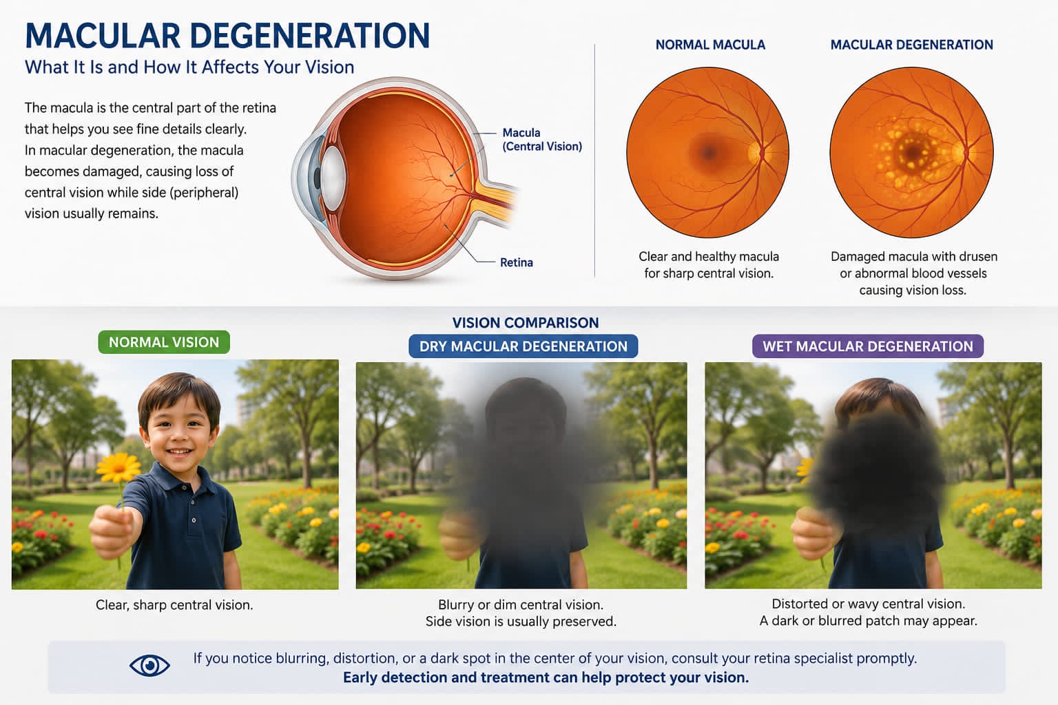
Macular degeneration is a condition that affects the macula, the central part of the retina responsible for sharp and detailed vision. The macula allows us to read, recognise faces, drive and see fine details clearly.
Macular degeneration most commonly occurs with increasing age and is therefore called Age-Related Macular Degeneration (AMD). It is an important cause of vision loss in people over the age of 50.
What Happens in Macular Degeneration?
The retina is the light-sensitive layer at the back of the eye. At the centre of the retina is a specialised area called the macula.
In macular degeneration, the macula gradually becomes damaged. This affects central vision, while peripheral or side vision is usually preserved. Patients may find it difficult to read or recognise faces, even though they can still see objects to the side.
Types of Macular Degeneration
There are two main types of age-related macular degeneration.
Dry Macular Degeneration
Dry AMD is the more common form. It occurs when the macula gradually becomes thin and degenerates with age.
- Small yellow deposits called drusen may develop beneath the retina.
- Dry AMD usually progresses slowly.
- In advanced stages, areas of the macula may become severely damaged, leading to significant central vision loss. This advanced form is known as geographic atrophy.
Wet Macular Degeneration
Wet AMD is less common but can cause rapid and severe loss of vision.
- Abnormal blood vessels grow beneath the retina and may leak fluid or blood.
- This can cause swelling and damage to the macula.
- Early diagnosis and treatment are extremely important in wet AMD because vision can deteriorate rapidly.
Symptoms of Macular Degeneration
Symptoms may include:
- Blurring of central vision
- Difficulty reading small print
- Straight lines appearing bent or wavy
- Difficulty recognising faces
- A dark or blurred patch in the centre of vision
- Colours appearing less bright
- Needing brighter light while reading
One important warning symptom is distortion of straight lines. Door frames, window grills or lines on a page may appear bent or wavy. If you notice sudden distortion or a sudden change in central vision, you should consult a retina specialist as early as possible.
Who Is at Risk?
The risk of macular degeneration increases with age. Other risk factors include smoking, a family history of AMD, high blood pressure, cardiovascular disease and certain genetic factors.
Smoking is one of the most important modifiable risk factors associated with macular degeneration.
How Is Macular Degeneration Diagnosed?
A detailed retinal examination is necessary to diagnose AMD.
The retina specialist may perform tests such as Optical Coherence Tomography (OCT). OCT provides detailed cross-sectional images of the retina and helps detect fluid, swelling and structural changes in the macula.
In some patients, additional retinal imaging or angiography may be required to study abnormal blood vessels beneath the retina.
Treatment of Dry Macular Degeneration
There is currently no single treatment that can completely reverse dry macular degeneration.
- Regular retinal examinations are important to monitor the condition and detect progression.
- In selected patients, specific combinations of vitamins and antioxidants may help reduce the risk of progression to advanced AMD. These supplements should be taken only after consultation with an ophthalmologist or retina specialist.
- Patients may also be advised to monitor their vision at home using an Amsler grid.
Treatment of Wet Macular Degeneration
Wet AMD is commonly treated with injections given into the eye.
- These medicines help reduce leakage from abnormal blood vessels and control swelling in the retina.
- Many patients require a series of injections and regular OCT scans to monitor the response to treatment.
Early treatment offers the best chance of preserving vision. Delaying treatment may result in permanent damage to the macula.
Can Macular Degeneration Cause Complete Blindness?
Macular degeneration mainly affects central vision. Even in advanced disease, peripheral vision is often preserved. Therefore, AMD usually does not cause complete darkness or total blindness.
However, severe central vision loss can significantly affect reading, driving and recognising faces. Low-vision aids and rehabilitation can help patients with advanced macular degeneration remain independent.
Can Macular Degeneration Be Prevented?
Not all cases of AMD can be prevented. However, certain lifestyle measures may help protect retinal health.
- Avoid smoking
- Maintain good blood pressure and cardiovascular health
- Eat a balanced diet rich in green leafy vegetables
- Attend regular eye examinations, particularly after the age of 50
Patients with AMD in one eye should have regular retinal examinations of both eyes.
When Should You See a Retina Specialist?
Consult a retina specialist if you notice sudden blurring of central vision, straight lines appearing bent or wavy, a dark patch in the centre of your vision or a sudden difficulty in reading or recognising faces.
Early detection and timely treatment are particularly important in wet macular degeneration.
If you have been diagnosed with macular degeneration or have noticed changes in your central vision, a detailed retinal examination and OCT scan can help identify the cause and guide appropriate treatment.